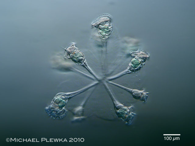
| Conochilus unicornis; a pelagic rotifer forming colonies. In this image the gelatinous sheath is visible. (2) |
| |
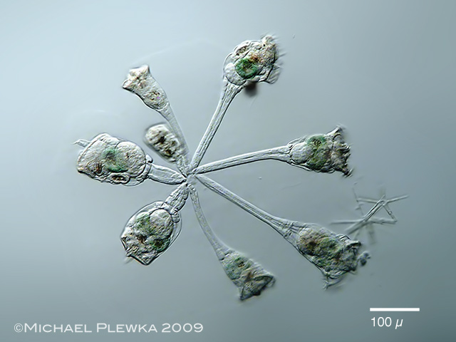 |
| Conochilus unicornis; specimen from (1) |
| |
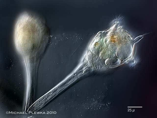 |
| Conochilus unicornis, showing the single (connate) lateral antenna of the right specimen. (2) |
| |
 |
| Conochilus unicornis, single individuum sorrounded by the mucilaginous sheath that keeps the colony together. (1) |
| |
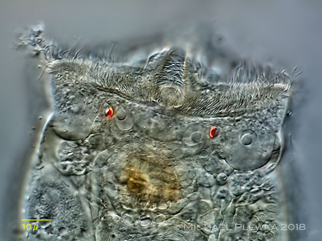 |
| Conochilus unicornis; this and the two images below show 3 aspects of the head reagion. Focus plane on the eyespots with a primitive lens on the dorsal fringe of the wheel organ. (3) |
| |
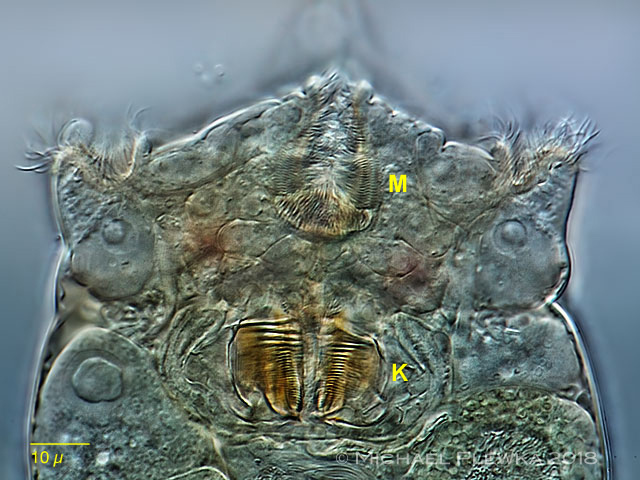 |
| Conochilus unicornis; 2nd aspect: focus plane on the mouth (m) and the trophi (K). (3) |
| |
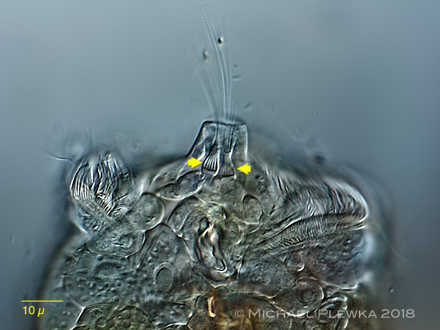 |
| Conochilus unicornis, 3rd aspect: focus plane on the ventral lateral antenna: in contrast to Conochilus hippocrepis the lateral antenna consists of two ciliated sensory cells (arrowheads) in a common housing. (3) |
|
| |
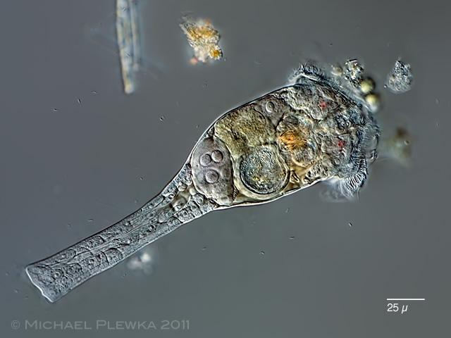 |
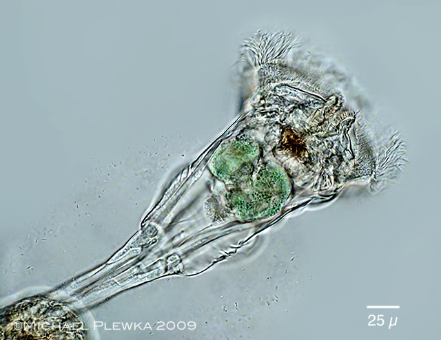
| Conochilus unicornis, single specimen (out of a colony) from (3) |
| |
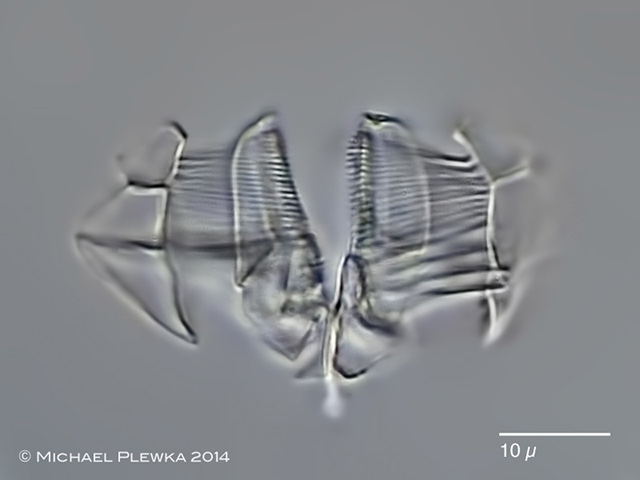 |
| Conochilus unicornis, malleoramate trophi (4) |
| |
| |
| |
| Location: Glörtalsperre / NRW (1); Heilenbecker Talsperre, NRW (2); Wahner Heide near Colgne (3); Hattingen, NRW, Ruhr, Teich (4) |
| Habitat: Plankton (1);(2); pond with Sphagnum (3); Plankton (4) |
| Date: 4.6.2009 (1); 15.04.2010 (4); 01.04.2011 (3); 17.04.2014 (4) |
|
|